See some images from the 2022 Nikon Small World photography contest
Existence is stunning at all scales, from big to modest. Occasionally that splendor is hid beneath literal scales.
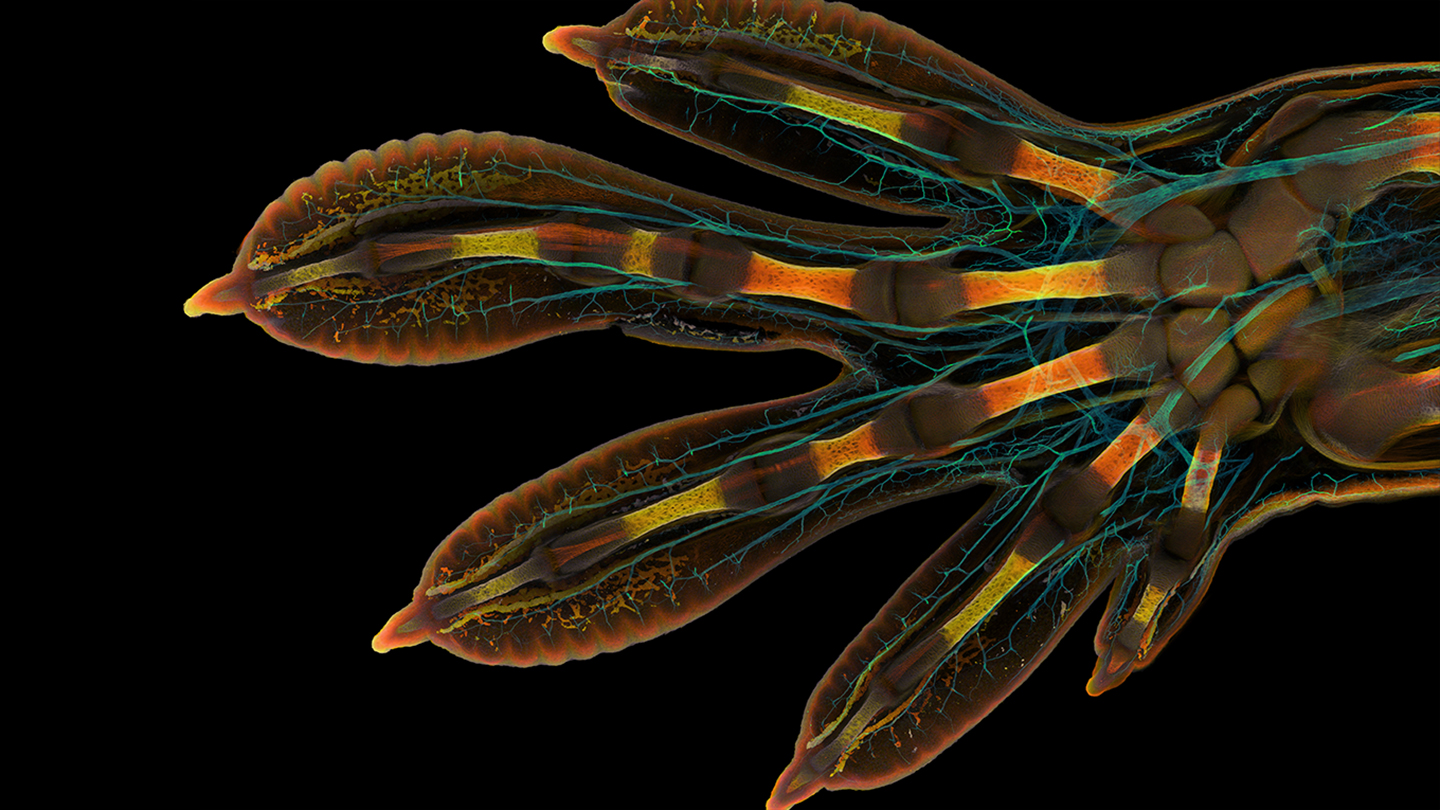
A mesmerizing peek beneath the creating scales on the hand of an embryonic Madagascar large working day gecko (Phelsuma grandis) gained to start with area in the 2022 Nikon Small Planet photomicrography level of competition. The successful impression, stitched jointly from hundreds of pictures taken more than two times with a confocal microscope, was crafted by College of Geneva researchers Grigorii Timin and Michel Milinkovitch. The pair analyze the genetics and physics of embryonic enhancement.
The hand is artificially coloured to demonstrate budding nerves in cyan and structures made up of collagen in a assortment of oranges and yellows. Collagen is a building block of everyday living, says Milinkovitch. Recognizing exactly where collagen is can assistance scientists greater realize how bodies and tissues produce.
Sections of bones that have started to calcify glow brightest in the graphic, Timin claims. Developing tendons and ligaments extend as orange branches. Blood cells type clusters or line up inside new blood vessels at the tips of the gecko’s digits.
The impression highlights attractiveness of all dimensions, Milinkovitch says. The snapshot is “beautiful as a hand, you see this lovely sample of the fingers. Then you zoom, you see the spongy bones. And you zoom, you see the tendons. And you zoom, and you see the fibers that is from the tendons. Then you zoom, and you see the blood cells.”
The gecko photograph is a single of 92 extraordinary photos acknowledged in this year’s level of competition. The winners of the 48th once-a-year contest had been announced Oct 11. In this article are some of our other favorites.
Bought milk?
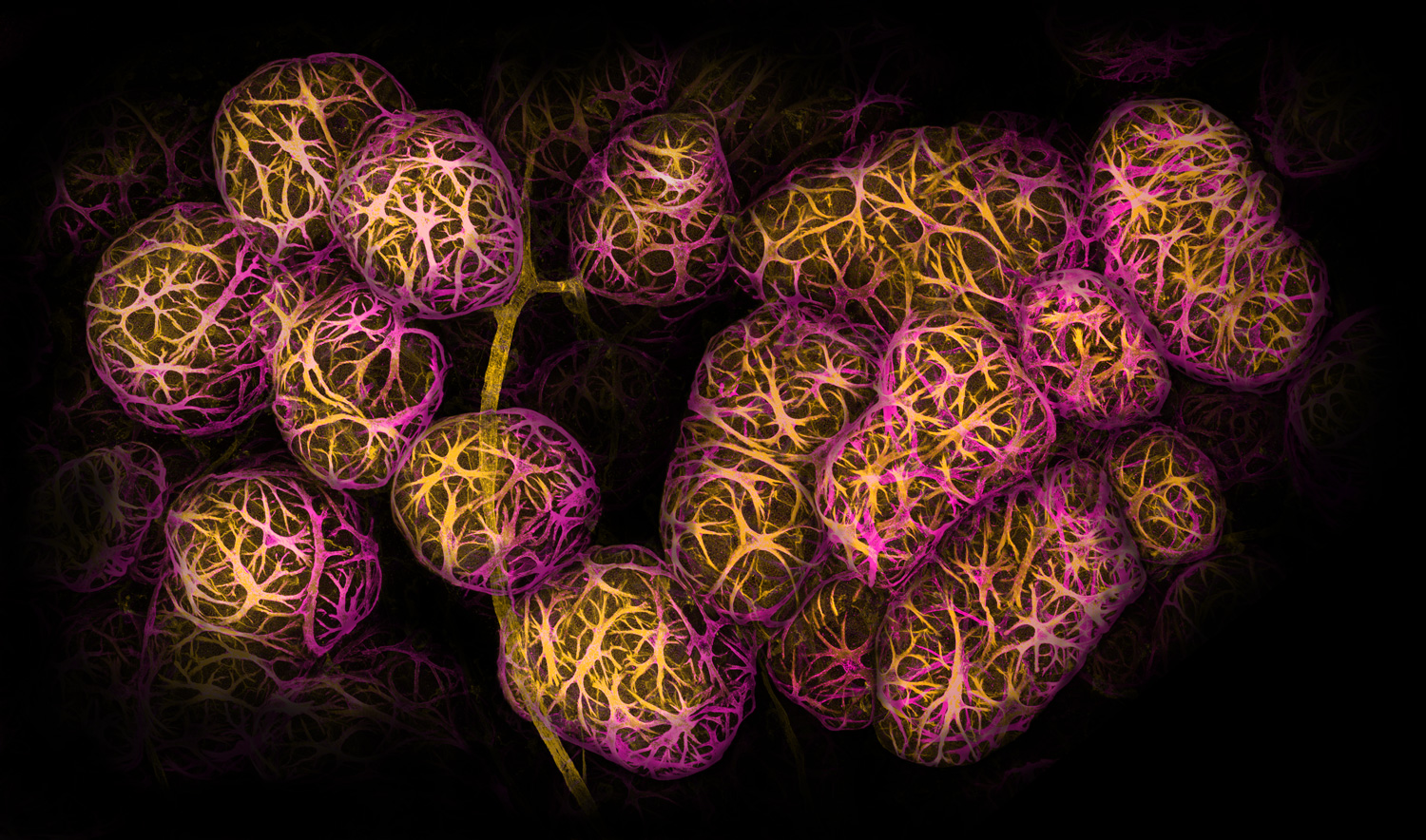
From a length this photograph may possibly seem like a cluster of grapes. But every single orb is a sprawling clump of cells inside breast tissue.
Most cancers immunologist Caleb Dawson of the Walter and Eliza Hall Institute of Health-related Analysis in Parkville, Australia, took countless numbers of illustrations or photos working with a confocal microscope to perspective little, musclelike cells that wrap around milk-developing spheres. He utilized dyes and antibodies to mark the cells yellow and magenta in this 2nd-position-successful impression.
The cells react to the hormone oxytocin, Dawson suggests. Oxytocin is unveiled during breastfeeding and can help squeeze milk out of the spheres, identified as alveoli. These types of illustrations or photos of lactating breast tissue can enable scientists figure out how immune cells retain breast tissue and the toddlers it can feed healthy.
Snuffed candle
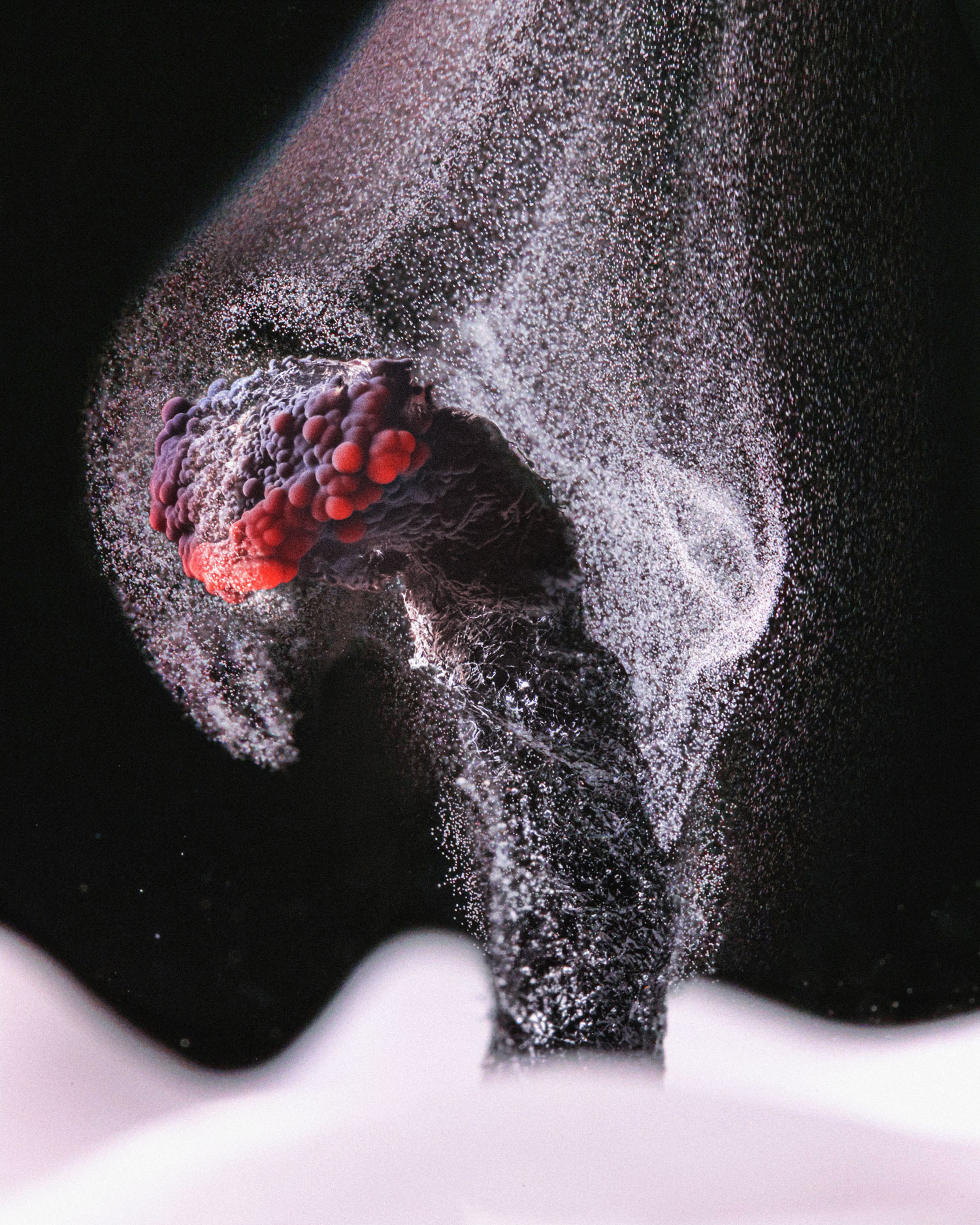
Ole Bielfeldt experienced to be quick to capture the very last gasp of an extinguished candle.
Candle wax is designed of hydrogen and carbon atoms, which turn mainly into carbon dioxide when lit on fireplace. But not all people hydrocarbons melt away, instead accumulating as soot on surfaces close to the candle. “When the flame goes out, the glowing wick has more than enough warmth still left to crack up the wax molecules for a although, but not plenty of to burn the carbon,” states Bielfeldt, a photographer in Cologne, Germany. “So you get a trail of smoke right until it cools.”
Working with a rapid shutter pace and a shiny LED gentle, Bielfeldt managed to seize all those unburned particles of carbon drifting absent, earning sixth location.
Iridescent slime mildew
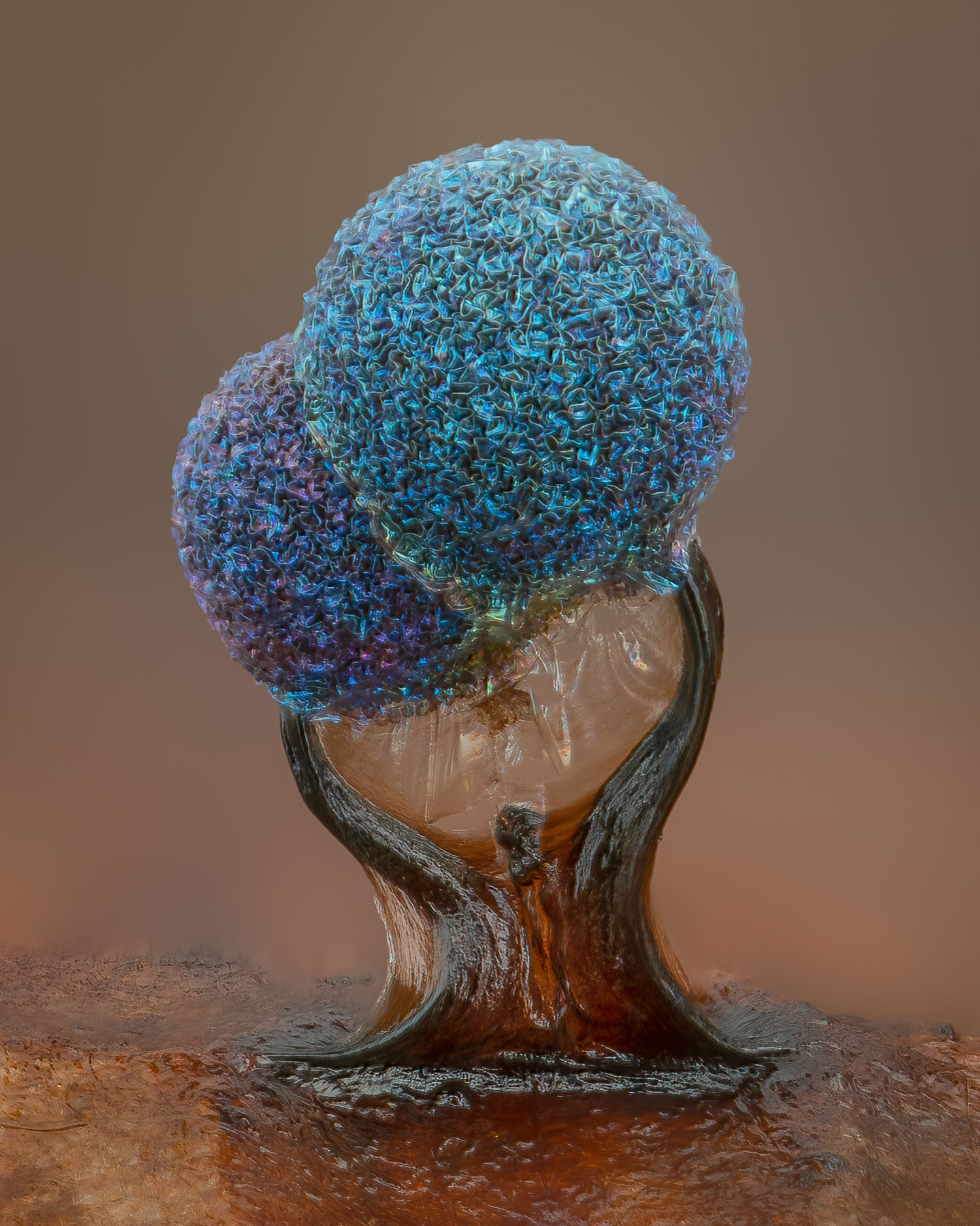
Hidden on leaves and decaying logs in moist forests are minuscule is effective of art like these Lamproderma slime molds.
In the dappled sunlight of an October working day, photographer Alison Pollack of San Anselmo, Calif., noticed a sparkly leaf as she was digging by way of a leaf pile. After using the leaf house and searching by way of a microscope, she was transfixed by the crinkly heads and iridescence of slime mould. About 40 hrs of do the job and 147 merged photographs later, Pollack experienced captured a hanging snapshot that she likes to anthropomorphize as a nurturing marriage: parent and child, two enthusiasts, or brother and sister. The photo gained fifth put in the contest.
Most slime molds have clean heads, which launch spores into the surroundings to reproduce. This pair might have dried as well fast, stunting their advancement and leaving their heads wrinkly, Pollack states. But that’s Ok, “because the texture to me is just gorgeous.”
A deadly predator
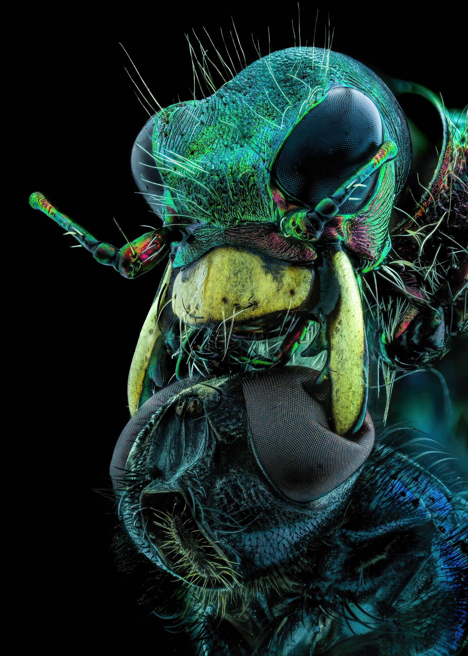
All fear the predacious tiger beetle, specially this very poor fly.
Murat Öztürk of Ankara, Turkey, nabbed 10th put in this year’s level of competition with an astounding — and unnerving — snapshot of a tiger beetle using its mandibles to crush a fly by its eyes.
Tiger beetles (Cicindelinae) sprint after their prey so rapidly that the bugs go briefly blind. The photographed beetle would have stopped various moments to orient alone to determine out wherever the fly was, ultimately snatching its food. Thanks to the beetle’s strong and sharp jaws, “the likelihood of survival of the creatures caught by this insect are incredibly reduced,” Öztürk suggests.
Coral near-up
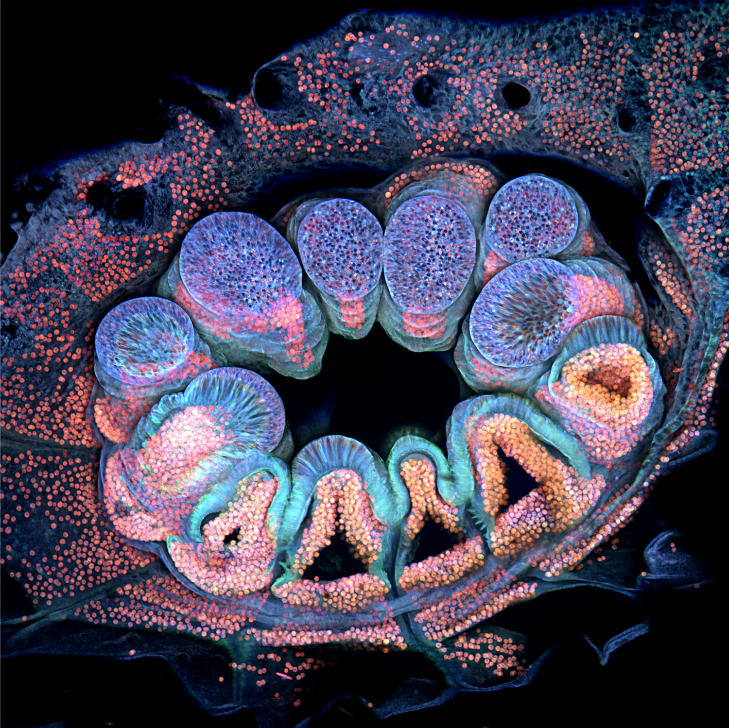
In Opal Reef off the coastline of Australia, some cauliflower coral (Pocillopora verrucosa) polyps surface inexperienced. But the same organism is remodeled when seen underneath a microscope in the lab.
To reveal the polyp’s particular person cells, maritime scientist Brett Lewis of Queensland College of Technology in Brisbane, Australia, stitched with each other extra than 60 photographs taken more than 36 several hours. The coral by natural means fluoresces a medley of blues, purples and pinks when uncovered to diverse wavelengths of gentle. Algae dwelling within the polyp surface orange or pink, whilst the coral’s tissues shine blue. The picture gained 12th spot in the competitors.
One particular wonderful point about the picture, Lewis states, is that in some parts, algal cells shine by way of a mild blue haze. Which is due to the fact coral tissue is transparent algae give coral its colour.
Peeks of coral’s internal makings can aid researchers understand its biology, Lewis says. His operate, for instance, aims to figure out how young polyps make solid foundations when they connect to a floor — an vital move in creating or restoring coral reefs.


